
NuDyRection
Nucleolar Dynamics during DNA Repair In Action
Project Research Axis 1
Axis 1 – How SMN Protein Moves and Maintains Nucleolar Health
Our team studies how the SMN protein, essential for cell function, moves between Cajal bodies and the nucleolus and helps maintain their proper organization. We have shown that after UV light or oxidative stress, SMN travels in a precisely timed way, guided by its partners Coilin (COIL), Fibrillarin (FBL), and PRMT1.
We are now exploring other proteins that may work with SMN, including newly discovered partners, which could play important roles in nucleolar organization and DNA repair. Using advanced BioID proteomics in engineered cells, we aim to map the network of proteins coordinating these movements and interactions.
This research helps us understand the complex protein network that keeps the nucleolus and Cajal bodies functioning, ensuring the cell can recover from DNA damage and maintain its health.
Techniques: BioID, immunoprecipitation (IP), proximity ligation assay (PLA), immunofluorescence (IF), Unscheduled DNA Synthesis (UDS), Transcription-Coupled Repair UDS (TCR-UDS), RNA Recovery Synthesis (RRS), RNA FISH, GST pull-down

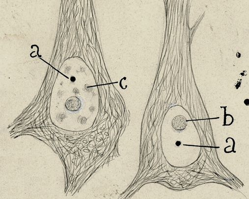
Santiago Ramón y Cajal – 1923.
Fig. 142. — Núcleo de las pirámides cerebrales del hombre. — a, cuerpo accesorio; b, nucleolo; c, grumos hialinos. Nótese que, usando ciertos fijadores, el proceder argéntico tiñe exclusivamente el cuerpo accesorio.
© Herederos de Santiago Ramón y Cajal
https://cvc.cervantes.es/ciencia/cajal/cajal_recuerdos/recuerdos/laminas.htm
Project Research Axis 2
Axis 2 – How Cajal Bodies and the Nucleolus Respond to DNA Damage
Our research explores how Cajal bodies and the nucleolus, important structures within the cell nucleus, change their organization when DNA is damaged and repaired. Using advanced 3D super-resolution microscopy (STED and STORM), we can see in detail how key proteins — SMN, Coilin, Fibrillarin (FBL), and RNAP1 — move and rearrange after stress such as UV light or oxidative damage.
By using live-cell imaging with fluorescently tagged proteins, we can watch these movements in real time, observing how SMN shuttles between Cajal bodies and nucleoli during DNA repair. We also use FRAP (Fluorescence Recovery After Photobleaching) to measure how fast these proteins move and how their interactions change during and after repair.
Together, these studies help us understand how the nucleolus and Cajal bodies adapt to DNA damage, ensuring the cell can recover and maintain its normal functions.
Techniques: Super-resolution microscopy (STED, STORM), live-cell imaging, FRAP, immunofluorescence
Project Research Axis 3
Axis 3 – Protecting SMN-Deficient Cells from Oxidative Stress
We study how cells lacking SMN respond to oxidative stress, which makes them more vulnerable to DNA damage and reduces their growth. The main problem is repairing oxidative DNA lesions, as key repair proteins cannot bind properly in SMN-deficient cells.
We investigate these repair processes in living cells and test whether antioxidants, such as N-acetyl-L-cysteine (NAC), can improve survival. We also examine how nerve injury and muscle denervation disrupt nucleoli in muscle cells and collaborate with SMA mouse model studies to better understand disease mechanisms.
This research aims to identify strategies to protect SMA cells and inform potential therapies.
Techniques: Clonogenic assays, immunofluorescence, chromatin fractionation, oxidative stress induction, antioxidant treatments.


Project Research Axis 4
Axis 4 – Studying SMN Function in Post-Mitotic Cells
Our research extends to more relevant cell types for SMA, including motor neurons and muscle cells, because SMA patients often show severe motor defects and congenital heart malformations.
We are testing whether SMN is essential for nucleolar organization in these cells and whether it moves within the nucleolus after DNA damage, as observed in fibroblasts.
To do this, we created human iPSC lines with inducible SMN depletion, allowing us to reduce SMN levels at specific stages of cell differentiation. Different clones have different residual SMN amounts, mimicking mild to severe SMA patients.
These models let us study how much SMN is needed at different developmental stages for proper cell function and survival. Importantly, all clones share the same genome, so differences are solely due to SMN levels.
Together, these new cell lines will complement existing SMA iPSC models and provide a more physiologically relevant system to validate SMN’s role in nucleolar function and DNA repair.